Exploring Veterinary Ophthalmoscope Use
News
Eye on Animal Health: Exploring Ophthalmoscopes in Veterinary Medicine
Ophthalmoscope veterinary use centers on examining the eyes of animals in detail, making ophthalmoscopes an essential piece of equipment for veterinarians, veterinary professionals, and clinics that need to diagnose eye conditions accurately. By allowing inspection of the retina, optic nerve, and other internal structures, these instruments help identify problems such as cataracts, glaucoma, retinal disease, and even tumors early, when treatment decisions can have the greatest impact on an animal’s vision, comfort, and overall health.
This guide focuses on how ophthalmoscopy supports veterinary diagnosis and monitoring, the types of ophthalmoscopes and specialized veterinary models available, the key features to consider when choosing one, how they are used in practice, and the veterinary ophthalmology products and services offered by Eickemeyer.

The Key Benefits of Ophthalmology and Complete Ophthalmic Examination in Veterinary Practice Include:
Early Detection: Ophthalmology allows for the early detection of eye diseases and conditions, which is crucial for timely intervention and treatment with medical technology.
Assessment of Retinal Health: The ophthalmoscope enables veterinarians to assess the health of the retina and optic nerve, which is essential for vision. This is especially important in animals with hereditary or acquired eye conditions.
Monitoring Progression: For animals with chronic eye conditions, regular ophthalmoscopy can help monitor the progression of the disease and the effectiveness of treatment.
Educational Tool: Ophthalmoscopy can be used as an educational tool to show clients the condition of their pet's eyes and explain the treatment plan.
Research and Documentation: Veterinarians use ophthalmoscopy to document the state of an animal's eye health, which can be valuable for research, user manuals, or legal purposes.
We Sell a Wide Range of Vet Equipment
Eickemeyer is a well-established and trusted supplier of veterinary equipment, known for its commitment to providing high-quality products and trustworthy services to the veterinary community. With a rich history spanning several decades, Eickemeyer has earned a strong reputation in the field of veterinary medicine for the following reasons:
Wide Range of Products: Eickemeyer offers an extensive range of veterinary equipment, covering various aspects of veterinary needs. From diagnostic tools to surgical instruments and equipment, we aim to meet the diverse needs of veterinarians and veterinary clinics.
Quality Assurance: Eickemeyer is dedicated to maintaining the highest standards of quality in its products. Veterinary professionals can trust that Eickemeyer's equipment is reliable, durable, and designed to meet the rigorous demands of veterinary care and improving medical technology.
Innovative Solutions: Eickemeyer consistently seeks to provide cutting-edge solutions for veterinarians. We stay up to date with medical technology advancements in the field and offer innovative products that enhance diagnostic accuracy and treatment outcomes.
Customer Support: Eickemeyer is committed to excellent customer service & communication. Our knowledgeable and friendly team is ready to assist with product inquiries, technical support, and any concerns that may arise using our products.
Training and Education: In addition to supplying equipment, Eickemeyer is excited to announce our 2024 lineup of Ophthalmology courses, Upcoming Vet Education Seminars and Conferences | Eickemeyer (eickemeyercanada.ca) to support veterinarians with their eye exams.
Community Engagement: Eickemeyer Canda is actively engaged with the veterinary community, participating in trade shows, conferences, and events to connect with practitioners and understand their evolving needs.
Long-Standing Reputation: Eickemeyer's reputation is built on years of consistent service and reliability. Many veterinary professionals trust Eickemeyer as a go-to source for their equipment needs now and in the future.
Eickemeyer serves as a reliable and valuable supplier of veterinary equipment for veterinarians and veterinary professionals in Canada and the United States. Our commitment to quality, innovation, and excellent customer support makes us an ideal partner for professionals in North American veterinary medicine to rely on Eickemeyer for:
Comprehensive Equipment: Eickemeyer offers a comprehensive selection of vet equipment, including diagnostic tools, surgical instruments, user manuals, and more, to meet the diverse needs of veterinary practices.
Quality Assurance: Professionals in both countries can trust Eickemeyer for equipment that meets the highest standards of quality, ensuring durability and reliability in their day-to-day work.
Long-Standing Reputation: Their reputation is built on years of consistent service and reliability, making them a trusted source for equipment among veterinary professionals in both Canada and the United States.
Understanding the Ophthalmoscope
An ophthalmoscope is a specialized medical instrument used in both human and animal healthcare to easily examine the internal structures of the eye, including the retina, optic nerve, blood vessels, and other important components. The instrument consists of a light source, a series of lenses, and a viewing window that allows veterinarians and medical professionals to illuminate and magnify the interior of the eye.
The critical role of veterinary ophthalmoscopes in animal healthcare can be summarized as follows:
Early Detection of Eye Conditions: complete eye examination / invaluable tools for the early detection and diagnosis of eye conditions in animals. They enable veterinarians to visualize abnormalities such as cataracts, glaucoma, retinal diseases, and tumours within the eye. Early diagnosis allows for prompt treatment and can often prevent further progression of the condition, preserving the animal's vision and comfort.
Monitoring Eye Health: Regular eye examinations are essential for monitoring the ongoing eye health of animals, particularly in breeds prone to hereditary eye conditions. By tracking changes over time, veterinarians can provide proactive care, adapting treatment plans and interventions as necessary to maintain the best possible eye health for their patients.
Assessing Overall Health: Beyond their role in eye health, they are also instrumental in assessing an animal's overall health. Many systemic diseases, such as diabetes and hypertension, can manifest with eye-related symptoms. Ophthalmoscopes enable veterinarians to spot these signs, which, in turn, leads to early detection and management of underlying health issues.
Enhancing Diagnostic Precision: In cases of eye injuries, trauma, or foreign body presence, ophthalmoscopes are vital for visualizing the extent of damage or identifying the exact location of foreign bodies within the eye. This precision aids veterinarians in making informed decisions about the most appropriate course of action for treatment, whether it involves surgery, medications, or other interventions.
Ophthalmoscopes are essential tools in veterinary practice, providing veterinarians with the ability to thoroughly assess and diagnose eye conditions, monitor eye health, identify systemic health issues, and enhance diagnostic accuracy. Their critical role in animal healthcare contributes to the overall well-being of animals by ensuring early intervention, timely treatment, and the preservation of their vision and comfort. It is a valuable diagnostic and monitoring tool in veterinary medicine for assessing and managing a wide range of eye conditions in animals. Here are some specific applications of ophthalmoscopy in diagnosing and monitoring eye conditions:
Retinal Diseases: Ophthalmoscopy allows veterinarians to examine the retina for signs of retinal diseases such as retinal degeneration, retinal lessions, detachment, or inflammation. This is crucial for diagnosing and monitoring conditions like progressive retinal atrophy (PRA) in dogs.
Cataracts: Cataracts are a common eye condition in animals, particularly in dogs. Veterinary ophthalmoscopy helps in visualizing the opacity of the lens, its location, and the extent of cataract formation.
Glaucoma: Ophthalmoscopy assists in diagnosing and monitoring glaucoma, a condition characterized by increased intraocular pressure. Veterinarians can observe changes in the optic nerve head and assess the severity of the disease.
Uveitis: In cases of uveitis or inflammation of the uvea (the middle layer of the eye), ophthalmoscopy can reveal changes in the iris, vitreous humor, and other structures within the eye.
Tumours and Masses: Ophthalmoscopy is used to identify the presence of tumours, growths, or masses within the eye. It helps in evaluating the location and size of these abnormalities.
Inherited Eye Conditions: For breeds with a higher risk of inherited eye conditions, such as progressive retinal atrophy in many dog breeds, ophthalmoscopy is essential for early diagnosis and to track the progression of these conditions.
Trauma Assessment: Ophthalmoscopy is used to assess eye injuries and the extent of damage, helping veterinarians make decisions about treatment and possible surgery.
Monitoring Treatment Progress: After the diagnosis of an eye condition, ophthalmoscopy is used to monitor the effectiveness of treatment. Improvement or deterioration in the eye's condition can be observed over time.
Educating Pet Owners: Ophthalmoscopy is a valuable tool for educating pet owners about their animal's eye health. Veterinarians can show clients the condition of their pet's eyes and explain treatment options.
Research and Documentation: Ophthalmoscopy is crucial for documenting the progression of eye diseases and for research purposes. It helps build a comprehensive medical history for the animal.
Overall, ophthalmoscopy is a fundamental procedure for veterinarians to diagnose, monitor, and manage eye conditions in animals. It aids in early detection, treatment planning, and ongoing assessment of eye health.
Veterinarians and veterinary professionals play crucial roles in using ophthalmoscopy equipment to maintain and assess the eye health of animals. Here's an overview of the vital roles they play:
Expertise in Eye Anatomy and Health: Veterinarians have a deep understanding of animal eye anatomy and the various conditions that can affect it. This knowledge is vital in correctly positioning and using the ophthalmoscope to examine the eye.
Skill in Handling Equipment: They are skilled in the proper use of ophthalmoscopes, which can be a delicate and sensitive instrument. Vet techs can be responsible for setting up and preparing the equipment for use.
Patient Handling: Veterinarians and vet techs are trained in handling animals during the examination. They ensure the animal remains calm and still, which is essential for accurate ophthalmoscopy.
Diagnosis and Interpretation: Veterinarians are responsible for interpreting the findings from ophthalmoscopy. They can diagnose various eye conditions based on what they observe, and this is critical for determining the right treatment plan.
Treatment Planning: After diagnosis, veterinarians decide on the most appropriate treatment or management plan. Vet techs may assist with administering medications or carrying out treatments prescribed by the vet.
Monitoring Progress: Both veterinarians and vet techs are involved in monitoring the progression of eye conditions in animals. Ongoing ophthalmoscopy examinations are essential for assessing the effectiveness of treatment and making adjustments as needed.
Client Education: Veterinarians and vet techs communicate with pet owners about the diagnosis, treatment options, and the importance of regular eye examinations. They help clients understand the significance of maintaining their pet's eye health.
Research and Documentation: They maintain thorough records of ophthalmoscopy findings and treatment plans, which are essential for research, legal purposes, and for tracking an animal's eye health history over time.
Emergency Care: In cases of eye emergencies or sudden trauma, veterinarians and vet techs are trained to use ophthalmoscopy to quickly assess the situation and make rapid decisions regarding immediate care.
Continuing Education: Keeping up with advancements in ophthalmoscopy and eye care is vital. Both veterinarians and vet techs engage in ongoing education to ensure they are using the latest techniques and equipment effectively.
Veterinarians and vet technicians are integral to the proper use of ophthalmoscopy equipment in veterinary practice. Their expertise, skill, and dedication to animal eye health are vital for diagnosing, treating, and monitoring eye conditions, ultimately ensuring the well-being of their animal patients.
How Ophthalmoscopes Work

An ophthalmoscope is a specialized medical instrument used to easily examine the interior structures of the eye, including the retina, optic nerve, and blood vessels. It consists of several fundamental components, each serving a specific purpose in the examination. Here are the key components of an ophthalmoscope:
Light Source: It is equipped with a light source, typically an adjustable, bright, white light, that is used to illuminate the interior of the eye. This light source is crucial for visualizing the eye's structures.
Power Source: Ophthalmoscopes can be powered by various sources, including batteries or wall outlets, depending on the model. A reliable power source is essential to ensure consistent illumination during examinations. Eickemeyer Canada has a Welch Allyn® Adaptor for Heine® Battery Handle available.
Aperture (Aperture Disc): The aperture is an adjustable opening or disc that controls the size and shape of the light beam. Different apertures are used for different types of examinations. Common apertures include:
Small Aperture: Used for a focused and narrow examination, ideal for assessing the macula and fine details of the retina.
Large Aperture: Provides a wider field of view and is used for the general examination of the fundus.
Slit Aperture: Creates a thin, vertical line of light, helpful for assessing retinal vessels and vitreous opacities.
Red-Free (Green) Filter Aperture: Filters out red light, enhancing the visualization of blood vessels and retinal details.
Diopter Adjustment: Ophthalmoscopes often have a diopter adjustment wheel that allows the examiner to focus the instrument for both the examiner's eyes and the patient's eye. This ensures a clear view of the eye's interior.
Lens System: Some ophthalmoscopes include a lens system that allows for magnification of the eye's structures. This is particularly useful for a more detailed examination of the retina and optic nerve.
Head and Handle: The head contains the light source, aperture, and lens system, while the handle houses the power source and controls. The handle is held by the examiner during the examination.
Eyepiece: Ophthalmoscopes have one or two eyepieces for the examiner to look through. In some models, the eyepieces can be adjusted to accommodate the examiner's eyesight.
Controls: Controls on the handle allow the examiner to adjust the intensity of the light, select different apertures, and manage other settings as needed during the examination.
Viewing Window: The viewing window is where the examiner looks to observe the interior of the eye. Some models include a built-in viewing window, while others may require the use of a separate viewing lens or indirect ophthalmoscope.
These fundamental components work together to provide veterinarians with the tools needed to perform detailed eye examinations and diagnose various eye conditions in animals.
Using an ophthalmoscope for eye examination in animals involves a systematic step-by-step process to ensure a thorough and accurate assessment of the eye's interior structures. Here's a detailed guide on how to use an ophthalmoscope for examination:
Room Lighting: Begin in a darkened room or an area with minimal ambient light. This allows for better visualization of the eye's interior.
Animal Restraint: Ensure that the animal is appropriately restrained to prevent sudden movements. Gentle restraint techniques may be necessary to keep the animal calm during the examination. Before viewing the fundus, examine the anterior segment, including pupil size and the anterior chamber for depth and transparency.
Use & Preparation:
Room Lighting: Begin in a darkened room or an area with minimal ambient light. This allows for better visualization of the eye's interior.
Animal Restraint: Ensure that the animal is appropriately restrained to prevent sudden movements. Gentle restraint techniques may be necessary to keep the animal calm during the examination.
Setup:
Power On: Turn on the ophthalmoscope and adjust the light source to an appropriate level for examination. Start with a low intensity and gradually increase it as needed.
Select Aperture: Choose the appropriate aperture for the specific type of examination you are conducting. Different apertures are used for different purposes (e.g., small aperture for macula examination, large aperture for general fundus examination).
Diopter Adjustment: Adjust the diopter settings to match your eyesight. Some ophthalmoscopes have a focus adjustment wheel to help achieve a clear image.
Approach to the Animal:
Position Yourself: Stand or sit at a comfortable distance from the animal, ensuring that you have a clear line of sight to the eye to be examined.
Examine One Eye at a Time: To minimize discomfort and stress for the animal, begin with one eye, and then move on to the other.
Examination Technique:
Illumination: Shine the ophthalmoscope light into the eye from a distance of about 15 inches (38 cm). Use your free hand to shield the eye not being examined, preventing excessive light from entering.
Approach and Angle: Approach the eye from the side, not head-on, to avoid direct light exposure. Hold the ophthalmoscope in your dominant hand, and use your other hand to gently hold the animal's head or eyelids, keeping them steady. Angle the ophthalmoscope slightly downward and aim it toward the center of the eye.
Start from a Distance: Initially, start your examination from a distance to get a general view of the fundus and optic nerve head.
Focus and Move Closer: Slowly move the ophthalmoscope closer to the eye while maintaining focus. As you approach the eye, you will be able to observe finer details.
Direct and Indirect Ophthalmoscopy: Depending on the type of ophthalmoscope being used, either look directly through the eyepiece of the ophthalmoscope or use an indirect ophthalmoscope with a separate viewing lens.
Examine the Fundus: Pay close attention to the retina, optic nerve, blood vessels, and other structures in the eye. Use the diopter adjustment to ensure clarity.
Move and Pan: Move the direct ophthalmoscope around to thoroughly examine the entire fundus, including the macula, retinal periphery, and any potential abnormalities.
Record Findings: Document any abnormalities or observations for future reference and for sharing with the animal's owner or for medical records.
Repeat for the Other Eye: Once the examination of one eye is complete, repeat the process for the other eye.
Power Off: Turn off the ophthalmoscope to conserve battery life.
Exit the Darkened Room: Gradually adjust the lighting back to normal if you are working in a darkened room.
Varieties of Ophthalmoscopes
Direct and indirect ophthalmoscopes are both valuable instruments for examining the interior structures of the eye, but they differ in their design, method of use, and the types of examinations they are best suited for. Here are the key differences between direct and indirect ophthalmoscopes:

Direct Ophthalmoscope
Heine® Beta 200 LED Ophthalmoscope Head - Eickemeyer Veterinary Technology (eickemeyercanada.ca):
Design: A direct ophthalmoscope is a handheld, compact instrument that combines the light source, aperture, and viewing optics in a single unit. It is relatively small and easy to handle.
Direct Viewing: The examiner looks directly through the eyepiece of the ophthalmoscope to observe the eye's interior. This direct viewing provides a real-time image of the fundus.
Pupil Dilation: Direct ophthalmoscopy typically requires that the pupil of the examined eye be dilated, either naturally through dim lighting or chemically with the use of dilating eye drops. This allows for better visualization of the fundus.
Field of View: The field of view is limited, which means that only a small portion of the fundus is visible at one time (fundus images). The examiner must physically move the instrument to explore the entire fundus.
Examination Distance: The direct ophthalmoscope is used at a close distance to the patient's eye, usually within a few inches.
Diagnostic Precision: It is highly precise for detailed examinations, especially when assessing specific areas of the retina, optic nerve, or macula. This makes it well-suited for pinpointing small lesions or abnormalities.
Commonly Used By: Direct ophthalmoscopes are often used by ophthalmologists and experienced clinicians due to the skill required to maintain proper alignment and focus during the examination.

Indirect Ophthalmoscope
HEINE® OMEGA 600 Indirect Ophthalmoscope - Eickemeyer Veterinary Technology (eickemeyercanada.ca):
Design: An indirect ophthalmoscope is a more complex and bulkier instrument that consists of a light source, a head-mounted lens, and a separate viewing lens. The examiner wears a headband with the lens apparatus.
Indirect Viewing: The examiner does not look directly through the ophthalmoscope. Instead, they use a separate viewing lens, which is placed a few inches from the patient's eye. This lens magnifies the image of the fundus, and the examiner views this magnified image through the lens.
Pupil Dilation: Pupil dilation is often not necessary for indirect ophthalmoscopy, making it a more practical choice for emergency or routine examinations.
Wide Field of View: Indirect ophthalmoscopy provides a wider field of view, allowing the examiner to see a larger portion of the fundus without moving the instrument. This is particularly useful for assessing the overall condition of the eye.
Examination Distance: The examiner maintains a greater distance from the patient's eye compared to direct ophthalmoscopy, making it more comfortable for both the examiner and the patient.
Diagnostic Precision: While not as precise as direct ophthalmoscopy for pinpointing small lesions, indirect ophthalmoscopy is excellent for surveying a broad area of the fundus. It is often used for initial assessments or emergency evaluations.
Commonly Used By: Indirect ophthalmoscopes are frequently used by general practitioners and in emergency departments due to their ease of use and the ability to quickly evaluate the fundus without pupil dilation.
The choice between direct and indirect ophthalmoscopy depends on the specific clinical situation and the examiner's skill level. Direct ophthalmoscopes provide high precision but may require more expertise and a dilated pupil, while indirect ophthalmoscopes offer a wider field of view and are often preferred for routine examinations or in situations where dilation is not practical.
Specialized Uses
Several specialized ophthalmoscopes are designed for specific veterinary applications, tailored to the unique needs of examining animals' eyes. These instruments cater to different animal species and clinical scenarios. Here are a few examples:
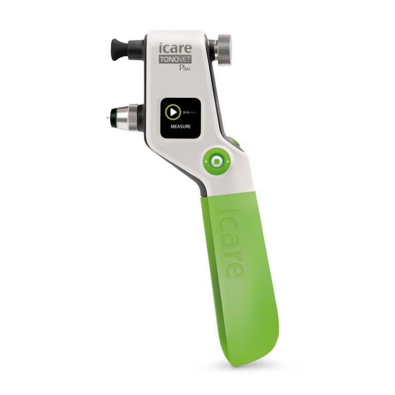
Vet: These ophthalmoscopes are specifically designed for veterinarians, allowing them to examine the eyes of various animals. They often come with a range of apertures and filters to facilitate detailed examination of different species. Icare® TONOVET Plus Tonometer - Eickemeyer Veterinary Technology (eickemeyercanada.ca)
Equine: Equine ophthalmoscopes are adapted for the larger size of horse eyes. They may have a longer reach to accommodate the anatomy of the horse's head.
Canine and Feline: These specialized ophthalmoscopes are optimized for examining the eyes of dogs and cats. They often come with smaller apertures and filters suited for small animal eye examinations.
Exotic Animal: Some veterinary ophthalmoscopes are designed for exotic animals, such as birds, reptiles, and small mammals. These instruments are adapted to the specific anatomical characteristics of these animals' eyes. Icare® TONOLAB Tonometer - Eickemeyer Veterinary Technology (eickemeyercanada.ca)
Portable Vet: These are lightweight and portable ophthalmoscopes designed for on-site examinations in field or farm settings. They are often battery-powered for convenience.


Digital Vet: Digital ophthalmoscopes allow for capturing images and videos of the eye, which is valuable for documentation, education, and telemedicine consultations in veterinary care. Slit Lamp Video Adaptor for Kowa SL-17 - Eickemeyer Veterinary Technology (eickemeyercanada.ca)
Fundus Cameras for Veterinary Use: While not traditional ophthalmoscopes, fundus cameras are specialized instruments for capturing high-resolution images of the retina in animals. These images can be valuable for diagnosing and monitoring eye conditions. ClearView® 2 Retinal Camera - Eickemeyer Veterinary Technology (eickemeyercanada.ca)
Equine Funduscope: Equine funduscopes are designed for imaging the retinas of horses and are particularly useful for diagnosing equine eye conditions.
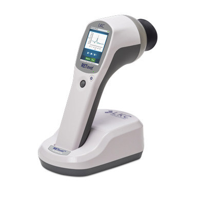
Canine and Feline Retinoscope: Retinoscopes are used to assess refractive errors in the eyes of dogs and cats. These specialized retinoscopes are designed for small animal ophthalmic examinations. RETevet Portable ERG - Eickemeyer Veterinary Technology (eickemeyercanada.ca)
These specialized ophthalmoscopes and related instruments are essential tools for veterinary ophthalmologists and clinicians, enabling them to provide precise and species-specific eye examinations, diagnoses, and treatments for a wide range of animals.
Key Features to Consider
Selecting the right ophthalmoscope is crucial for veterinarians, as it directly impacts their ability to diagnose and monitor eye conditions in animals. Here are some essential features that veterinarians should keep in mind when choosing an ophthalmoscope, along with the importance of quality and reliability:
Optical Quality: Optics are fundamental to the performance of an ophthalmoscope. Veterinarians should look for an instrument with high-quality lenses and optics to ensure a clear and sharp view of the eye's interior. Poor optics can lead to inaccurate diagnoses.
Illumination: Adequate and adjustable illumination is critical. The ophthalmoscope should offer a range of light-intensity settings to adapt to various clinical scenarios and animal species. Proper illumination is necessary for a detailed examination.
Apertures and Filters: Ophthalmoscopes should include a variety of apertures and filters to suit different types of examinations. Small apertures are essential for focused inspection of specific areas, while filters like red-free/green can enhance the visibility of blood vessels and retinal structures.
Magnification: Some ophthalmoscopes have a built-in lens system for magnification. This can be beneficial for examining small details in the retina but is not always necessary.
Diopter Adjustment: The ability to adjust the diopter settings is important for maintaining focus and clarity according to the examiner's eyesight. A diopter adjustment is particularly useful for clinicians who require corrective lenses.
Power Source: Consider whether the ophthalmoscope is battery-powered or powered by an electrical source. Battery-powered options provide greater mobility, while wall-powered ophthalmoscopes ensure continuous operation.
Portability: For fieldwork and ambulatory veterinarians, a portable ophthalmoscope can be advantageous. These models are lightweight and easy to carry.
Durability: Ophthalmoscopes should be built to withstand regular use and minor bumps. Choose a model with a robust construction that can handle the demands of a veterinary clinic or field setting.
Ease of Use: The instrument should be user-friendly, with intuitive controls and a comfortable grip. Veterinarians may have to perform lengthy examinations, so ergonomics are important.
Compatibility with Accessories: Ensure that the ophthalmoscope is compatible with various accessories, such as a slit lamp or a digital camera, for comprehensive examinations and documentation.
Animal-Specific Models: Depending on the variety of animals seen in the practice, veterinarians may opt for specialized ophthalmoscopes designed for certain species, like equine, canine, or feline models.
The importance of quality and reliability in veterinary equipment cannot be overstated. High-quality ophthalmoscopes are not only more precise and accurate in their examination but also tend to be more durable, ensuring a longer lifespan and fewer maintenance issues. Reliability is crucial in veterinary practice, as the instrument must perform consistently during crucial diagnoses and treatments.
Investing in reliable and high-quality equipment ultimately contributes to improved patient care, accurate diagnoses, and the overall success of the veterinary practice. Veterinarians should prioritize these features when selecting an ophthalmoscope to ensure it meets the specific needs of their practice and patients.
Eickemeyer Ophthalmoscope Products
Eickemeyer Canada has a vast selection of veterinary-specific ophthalmology products and supplies that range from surgical tools to diagnostic equipment and light sources.
Heine® BETA 200 LED Set 1 - Eickemeyer Veterinary Technology (eickemeyercanada.ca)
Heine® Finoff Transillumintor Head LED - Eickemeyer Veterinary Technology (eickemeyercanada.ca)
Heine® HSL 150 Set with Rechargeable Handle - Eickemeyer Veterinary Technology (eickemeyercanada.ca)
Heine®HSL 10x Converter Attachment - Eickemeyer Veterinary Technology (eickemeyercanada.ca)
Heine®Mini 3000 2.5V Ophthalmoscope - Eickemeyer Veterinary Technology (eickemeyercanada.ca)
Heine®Mini 3000 2.5V Ophthalmoscope Head - Eickemeyer Veterinary Technology (eickemeyercanada.ca)
HEINE® OMEGA Omega 600 Traveller Set - Eickemeyer Veterinary Technology (eickemeyercanada.ca)
Icare® TONOVET Plus Tonometer - Eickemeyer Veterinary Technology (eickemeyercanada.ca)
Why Choose Eickemeyer?
Veterinarians choose Eickemeyer for ophthalmology products because of our reputation for quality within the veterinary business, wide product range, commitment to customer support, and specialized focus on veterinary equipment. Eickemeyer's dedication to the well-being of animals and the success of veterinary practitioners makes us a preferred supplier of ophthalmology products in the veterinary field.
We encourage veterinarians and those in the veterinary business to explore Eickemeyer Canada's vast selection of vet equipment. Check out our supply of Ophthalmology products in our online catalogue Shop Online for Veterinary Equipment and Supplies - Eickemeyer (eickemeyercanada.ca)
Additional Resources
Ophthalmology_Catalogue_215,9x279,4mm_oP_Bro_32_CAN_engl_07-2020.indd (eickemeyercanada.ca)
Shop Ophthalmology - Eickemeyer Veterinary Technology (eickemeyercanada.ca)
Email info@eickemeyervet.ca or call us at 1 519 273 5558 to learn more about our vet products and shipping. Or check out our website Eickemeyer Canada for all your veterinary equipment needs.